- ABOUT JNU
- ADMISSION
-
ACADEMICS
- Schools and Colleges
-
Departments and Programs
- Arts College of
- Chinese Language and Culture College of
- Economics College of
- Electrical and Information Engineering College of
- Foreign Studies College of
- Information Science and Technology College of
- Environment School of
- Humanities School of
- International Business School
- International Studies School of
- Journalism and Communication College of
- Law School
- Liberal Arts College of
- Life Science and Technology College of
- Management School of
- Marxism School of
- Medicine School of
- Pharmacy College of
- Physical Education School of
- Science and Engineering College of
- Shenzhen Tourism College
- Research Institute
- Research Center
- Programs in English
- Majors
- Study Abroad
- Online Learning
- RESEARCH
- CAMPUS LIFE
- JOIN US
Latest News
Breakthrough in Neuroscience Imaging: JNU Team Unveils Revolutionary Fiber Optic Photoacoustic Microscope for Small Animal Brain Studies
Publisher: School of Physics and Optoelectronic Engineering
Date: January 11, 2024
In a momentous stride towards advancing the frontiers of neuroscience research, the pioneering team led by Professor Guan Baoou from the esteemed School of Physics and Optoelectronic Engineering at JNU has unveiled a groundbreaking achievement in the realm of small animal brain imaging. This monumental breakthrough, highlighted in the prestigious journal Light: Science & Applications, heralds a new era of innovation in the field of neuroimaging and critical care medical research.

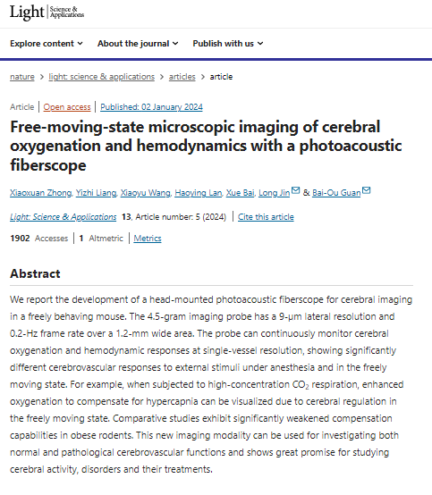
(Screenshot of the paper)
The transformative brain imaging technology developed by Professor Guan Baoou's visionary team represents a paradigm shift in the realm of small animal neuroscience studies, enabling real-time imaging of brain function in freely moving subjects. With unprecedented single-vessel resolution capabilities, this cutting-edge head-mounted fiber optic photoacoustic microscope offers a window into the dynamic oxygenation function and hemodynamic changes within the cerebral cortex layer, unlocking novel insights that hold profound implications for neuroscience and critical care medicine.
The seminal research findings, encapsulated in the paper titled Free Moving State Microsurgical Imaging of Cerebral Oxygenation and Hemodynamics with a Photoacoustic Fiber Scope, were unveiled on January 2, 2024, in Light: Science & Applications, a renowned publication renowned for its impact factor of 19.4.
Distinguished by its technical prowess and innovative design, the fiber optic photoacoustic microscope featured in this groundbreaking study boasts a compact form factor, exceptional sensitivity, unparalleled resolution, and versatile imaging modalities, catering to the unique demands of neuroscience and medical research. The adoption of this cutting-edge technology by emergency and critical care medical research teams promises to illuminate the intricate mechanisms underlying brain function impairment triggered by various diseases, laying the foundation for the precise formulation of targeted treatment strategies.
Moreover, the versatility of this state-of-the-art microscope extends beyond standalone applications, with the potential for seamless integration with complementary imaging modalities such as two-photon microscopy and wide-field fluorescence microscopy. This synergistic approach holds promise for unraveling complex physiological phenomena such as neurovascular coupling and delving into the underlying mechanisms of brain damage associated with neurodegenerative disorders like Alzheimer's disease.
This groundbreaking work, independently spearheaded by the trailblazing researchers at JNU, features Associate Professors Liang Yizhi and Dr. Zhong Xiaoxuan as co-first authors, alongside the esteemed Professors Guan Baoou and Jin Long as co-corresponding authors. The research endeavor received vital support from esteemed funding bodies, including the National Natural Science Foundation of China and the Guangdong Special Support Program Local Team Project.
For a comprehensive exploration of this transformative research initiative, readers are encouraged to delve into the original paper available at: [Paper Link] (https://www.nature.com/articles/s41377-023-01348-3).
NEWS
- About the University
- Quick Links
Copyright © 2016 Jinan University. All Rights Reserved.


